Independent study & small groups
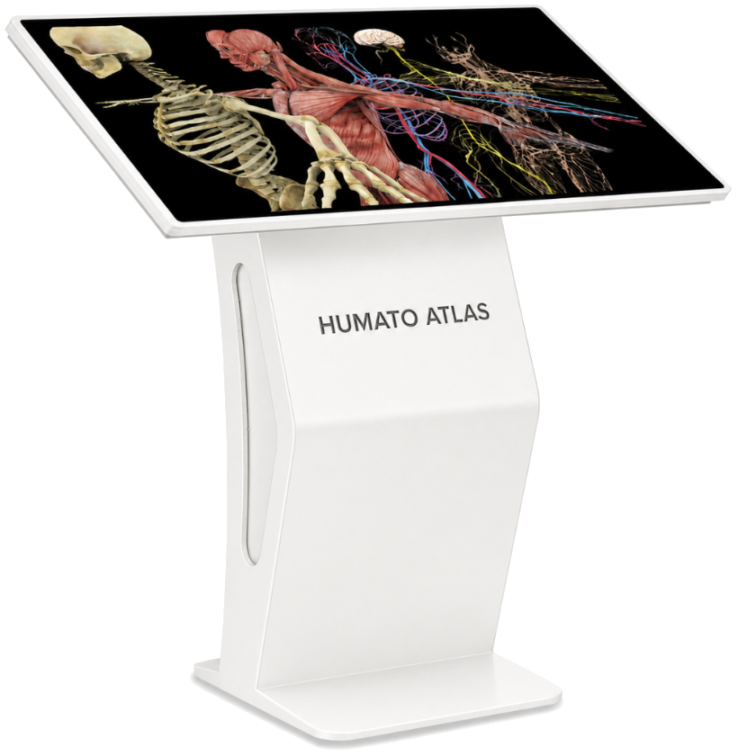
ATLAS MINI
Seminars, libraries & skills labs
The interactive anatomy table
Explore the body. Inspire the room.
Bring anatomical discovery, clinical understanding and shared discussion onto one teaching surface.
Room planning, faculty training and continuing support.
Choose a view
Inside Humato ATLAS
Explore, explain and discuss together.
 PATIENT’S RIGHTPATIENT’S LEFT
LANDMARK 01
Forms much of the anterior surface. Find it to the patient’s right of the anterior interventricular groove.
Why a table
A shared surface gives faculty and students a common reference for practical teaching. Isolate a structure, reveal its neighbours and relate the anatomy to a clinical image.
Alongside cadaveric teaching, virtual dissections can be repeated and cuts reversed. Restore a layer, revisit a difficult relationship and invite students to explain what they see.
See the teaching toolsInside the learning platform
From a structure to a scan, from tissue to movement. Explore the tools that help you explain the relationship, then let the class investigate.
Anatomy / teaching with ATLAS
Make a difficult relationship easier to explain. Isolate a structure, reveal its neighbours and return to the whole body as the lesson develops.
Explore · Isolate · Connect
Explore anatomyDisease & development / teaching with ATLAS
Compare healthy anatomy with animated disease progression. Follow development in maternal context, then compare singleton and twin pregnancy to explain the changing relationships.
Disease progression · Healthy comparison · Development
Explore disease & developmentSports anatomy / teaching with ATLAS
Slow the motion. Follow a muscle through an action and connect joint mechanics to function, load and injury.
Variable speed · Regional movements · Injury context
Explore sports anatomyDental anatomy / teaching with ATLAS
Move from the whole dentition to enamel, dentine and pulp. Compare disease stages, follow root canal treatment sequences and discuss prosthetic outcomes.
Tooth layers · Treatment sequences · Restoration
Explore dental anatomyDissection / teaching with ATLAS
Prepare students for the practical and help them revisit it afterwards. Narrated dissection connects the digital model to regional anatomy in the specimen.
Preparation · Narrated dissection · Revision
Explore dissectionHistology library / teaching with ATLAS
Navigate whole-slide scans, zoom into cellular detail and annotate the features that matter to the lesson.
Whole-slide scans · Cellular zoom · Annotation
Explore histology libraryClinical context / teaching with ATLAS
Move beyond naming a structure. Review a case and its images, discuss the findings and bring the class back to the anatomy that explains them.
Case discussion · Imaging · Clinical correlation
Explore clinical contextDICOM viewer / teaching with ATLAS
Import CT and MRI studies, reconstruct 2D slices into 3D and connect sectional findings with spatial anatomy. Compare views with synchronised scrolling and measurement.
CT & MRI · 3D reconstruction · Your studies
Explore DICOM viewerAssessment / teaching with ATLAS
Choose a topic, reveal answers together and review performance reports. Use answer feedback to decide which relationships need another explanation.
Answer feedback · Performance reports · Review
Explore assessmentFind your teaching format
Focused revision, a shared practical or a larger teaching group. Choose the surface around the lesson and the space.
Seminars, libraries & skills labs
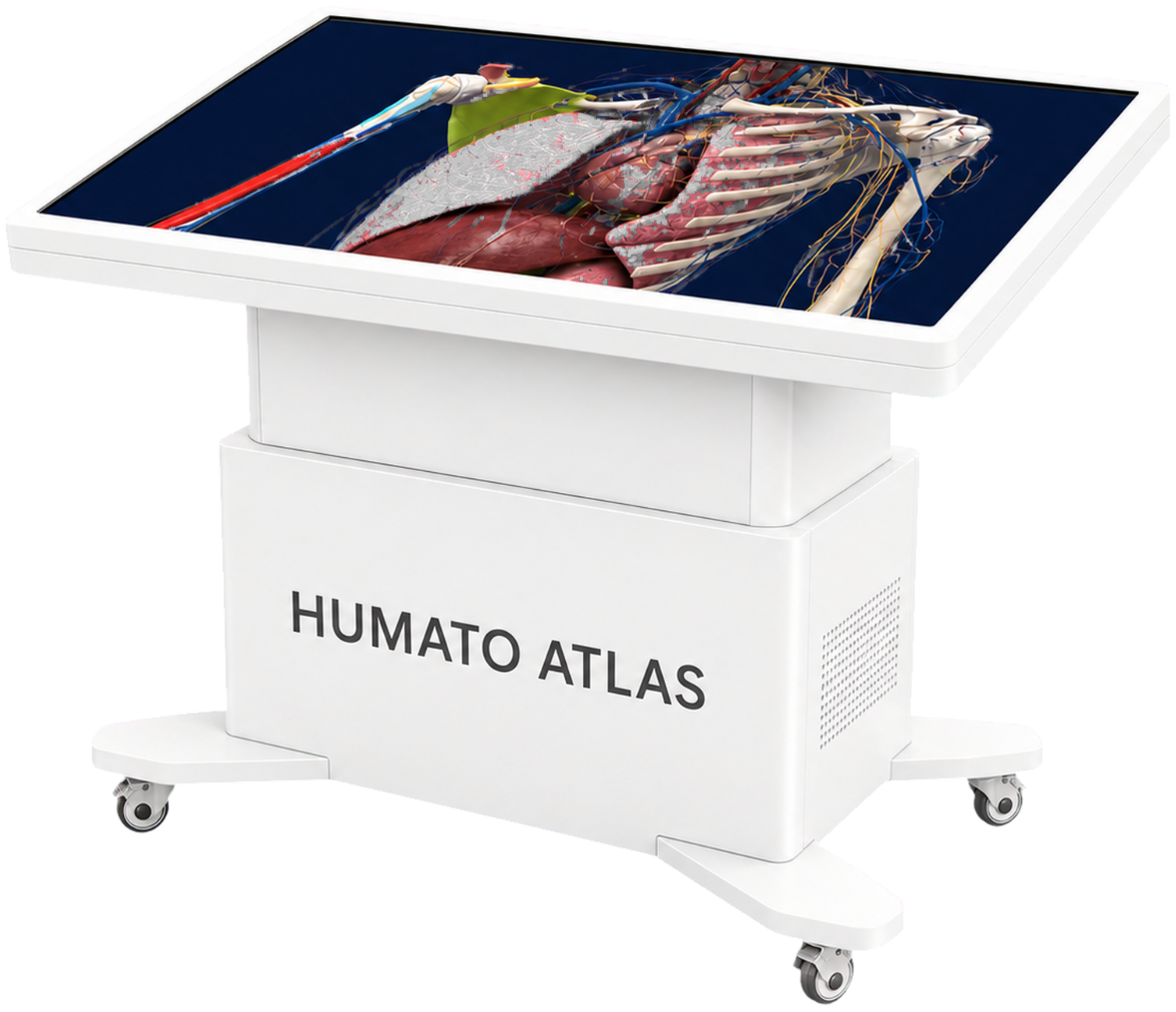
Dissection halls & tutorial rooms
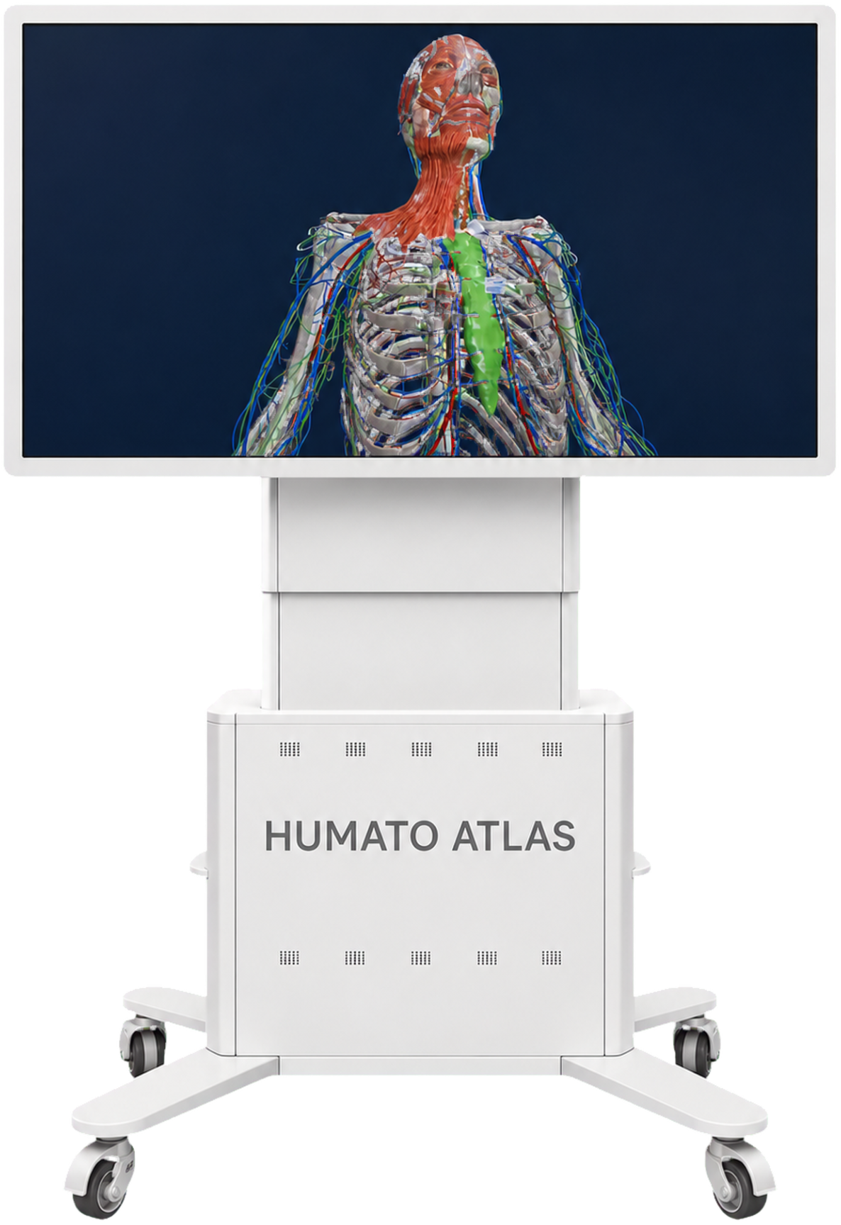
Anatomy labs & lecture rooms
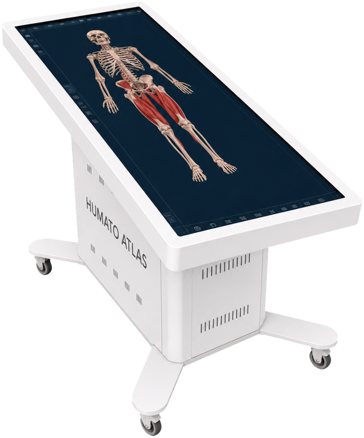
Dissection halls & anatomy museums
From selection to teaching
Work through the room, configuration and faculty needs before installation. Know what your proposed supply includes.
Compare formats around your course, group and available space.
Complete commissioning, acceptance testing and a documented handover, with faculty training around your curriculum.
Confirm software updates, warranty and maintenance within the agreed support terms.
Experience Humato ATLAS
Choose an anatomical relationship your students find difficult. Explore how you could explain it with ATLAS.